|
|
|
|
|
|
|
Parts of Lower Extremity.—The lower extremity consists of three parts, the thigh, the leg and the foot, and is united to the trunk by the os innominatum or haunch bone, which bears the same relation to the lower extremity that the bones of the shoulder do to the upper extremity.
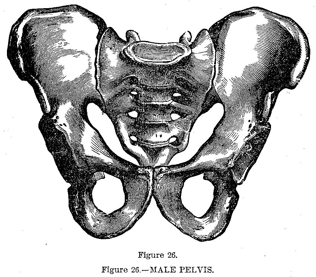
The os innominatum (Fig. 26) consists of the ilium, ischium and pubes, which in. the adult grow together and form one single bone. It is irregularly oblong in shape and twisted upon itself. The ilium is the broad upper part of the bone and forms the prominence of the hip. The ischium is the V-shaped lower portion upon which we sit. The pubes is situated in front and is also V-shaped; in the adult the upper part is covered by hair. Between these V-shaped bones is a large opening, the obturator or thyroid foramen.
Hip Socket.—At the junction of the three bones is a cup-shaped cavity, the acetabulum or socket of the hip, which receives the rounded head of the thigh bone. In front the pubic bones join and behind the sacrum complete the bony ring of the pelvis. The pelvis (Fig. 26) is basin-shaped, supports the contents of the abdomen and the trunk upon the limbs.
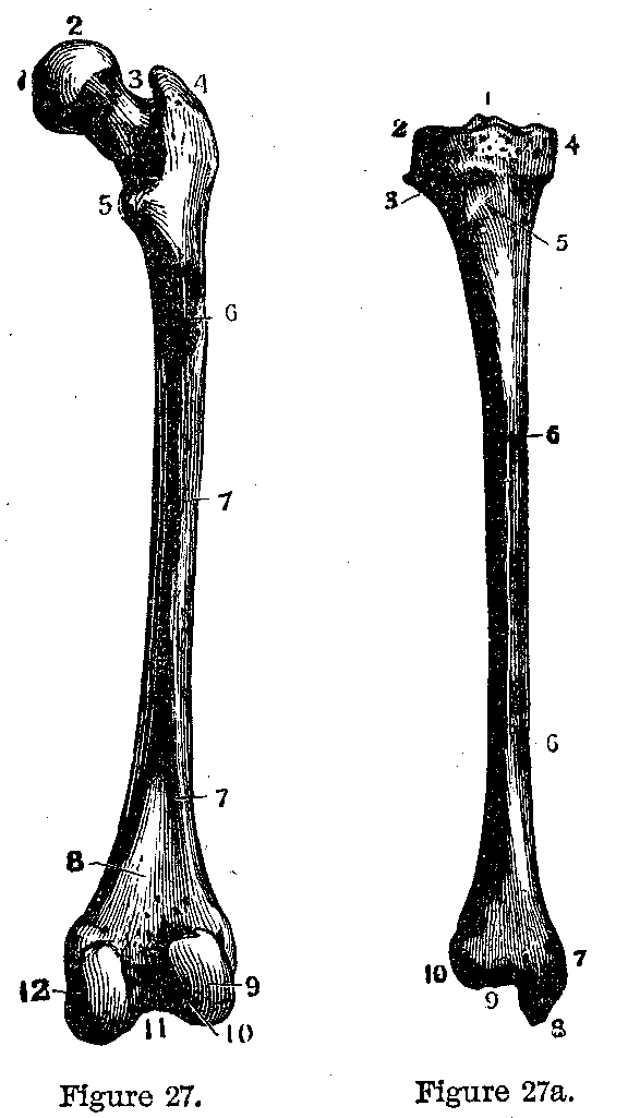
Thigh Bone.—The femur (Fig. 27) or thigh bone is the largest and strongest bone in the body. It consists of a shaft and two extremities. The upper extremity consists of a head which is spherical and smooth, fitting into the acetabulum, and a neck which joins the shaft at an obtuse angle. The shaft supports the body, is an important lever in locomotion and gives attachment to muscles. The lower extremity resembles the lower end of the humerus; it is smooth and joins with the main bone of the leg, the tibia.
Knee Pan.—The patella or knee pan is a small flat bone situated in the huge tendon of the great muscles on the front of the thigh. It protects the knee joint and increases the leverage.
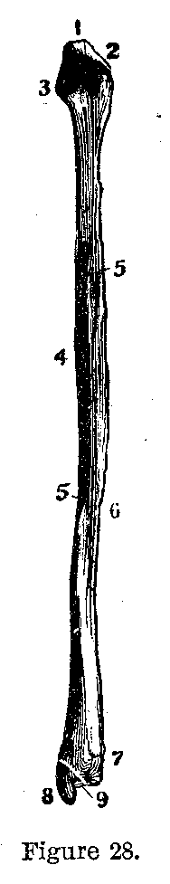
Leg Bones.—The leg bones are the tibia or shin bone (Fig. 27a) and the fibula (Fig. 28). The tibia, the larger and stronger, is expanded above to join the femur; the shaft is triangular, the sharp edge in front may be readily felt beneath the skin as the shin. The lower extremity forms the inner part of the ankle joint. The fibula (Fig. 28) is a long, slender bone lying on the outside of the leg. Its upper end joins the expanded upper extremity of the tibia, strengthening it, the lower end forms the outer part of the ankle joint.
|
|
|
|
|
|
|
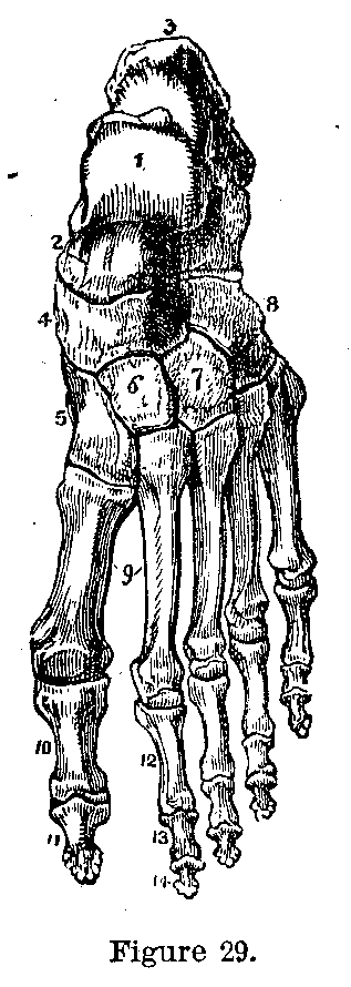
Foot Bones.—The foot (Fig. 29) consists of the tarsus or ankle bones, the metatarsus or foot bones and the phalanges or toe bones. The bones of the tarsus are the calcaneum, os calcis, or heel bone, the astragulus which joins the bones of the leg, the cuboid, the scaphoid and the three cuneiform bones. There are five metatarsal bones corresponding to the metacarpal bones of the hand. The phalanges are similar to those of the hand, there being two for the great toe and three for each of the other toes.
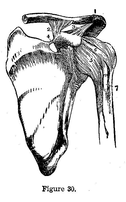
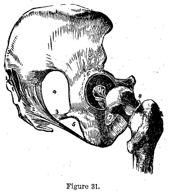
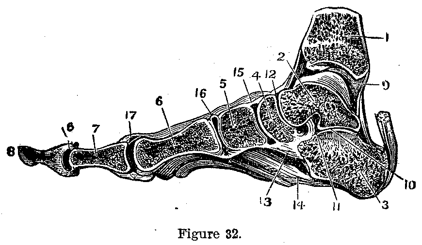
Where two bones meet a joint or articulation exists. The bones may be so soldered together (Fig. 30) as to form an immovable joint, as in the bones of the skull; they may be slightly movable as the pelvic and vertebral joints, or they may be freely movable as in most of the articulations of the limbs. The freely movable joints (Fig. 31) are the hinges, as the elbow, the ball and socket, as the shoulder, the gliding, as the sterno, clavicular articulation, and the ring and pivot (Fig. 32) joint, as the atloaxoid articulation. The structures entering into joint formation are bones, cartilages, ligaments and synovial membrane which secretes the lubricating fluid of the joint. (Figs. 30, 31 and 32.)
|
|
|
|
|
|
|
It will be unnecessary to describe the individual joints, reference having already been made to them in the section on bones.
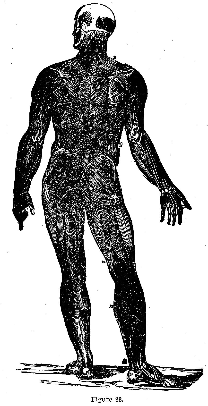
Function of Muscles.—Muscles are familiar as the flesh of animals. They are attached to bones, ligaments, cartilages and the skin, and by their contractions cause all the movements of the body. Some muscles are arranged in sheets (Fig. 33), some are spindle-shaped, some are disposed in rings like the muscle which closes the mouth, and in some the fibres spread out like a fan.
|
|
|
|
|
|
|
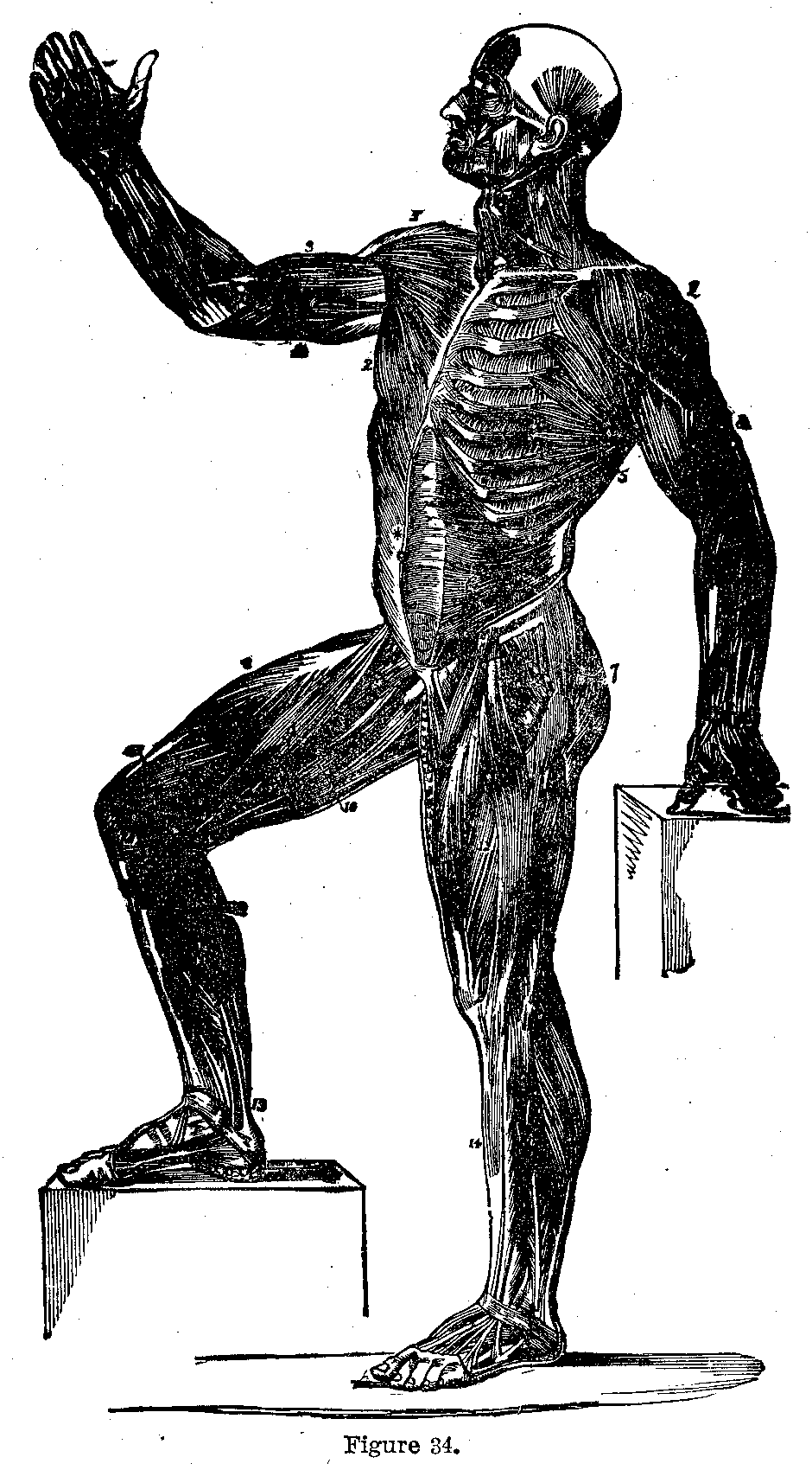
Muscle Attachments.—They are attached by fibrous cords, the tendons, or by broad fibrous bands, the aponeuroses. The end of the muscle which has the firmer attachment is called its origin (Fig. 34), the other end its insertion; this is, as a rule, merely relative, as in most cases the muscles act from either extremity; for instance, the sterno-cleido mastoid, the muscle which forms the prominent cord at either side of the neck, has its origin from the top of the breast bone and the end of the collar bone, and its insertion into the bony prominence of the skull behind the ear, its action is to bow the head and turn the face to the opposite side; but if the head be fixed it serves to raise the ribs and is thus an accessory muscle of respiration.
|
|
|
|
|
|
|
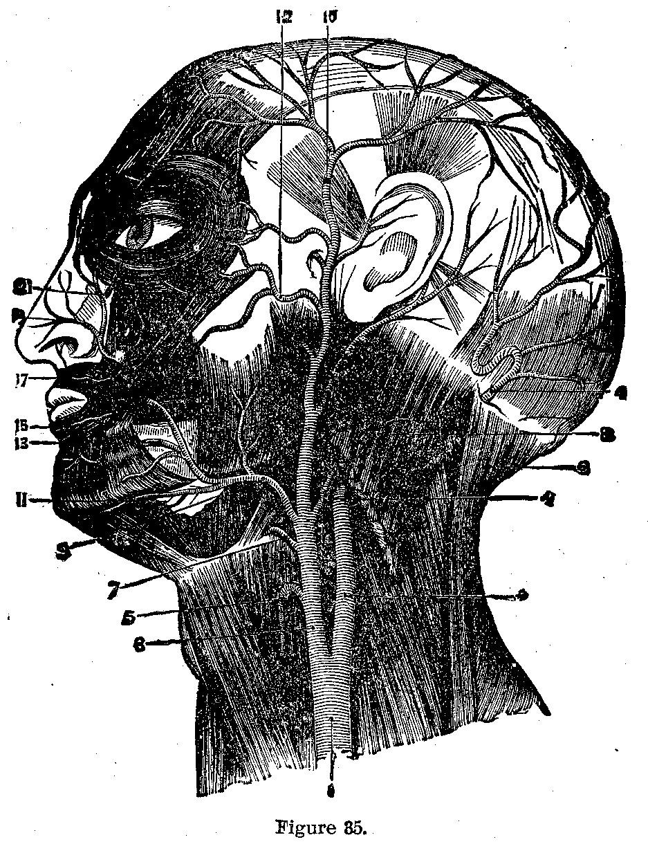
Face Muscles.—Of the numerous small muscles of the face (Fig. 35), it is not necessary to speak here; as a rule they arise from the bones of the face and are inserted into the skin, by their mobility giving expression to the countenance.
Muscles of Chewing.—The muscles of the orbit will be taken up in connection with the eye. The muscles of mastication are the temporal, masseter, the two pterygoids and the buccinator. The temporal arises from the side of the head above the ear and is inserted into the top of the lower jaw. The masseter runs from the bony process external to the orbit, to the angle of the jaw and forms the hard mass felt in the cheek when the Jaw is tightly closed.
|
|
|
|
|
|
|
Muscles of the Jaw.—The pterygoids run from the base of the skull to the lower jaw, moving it laterally. The buccinator is a broad, flat sheet in the cheek compressing the cheeks as when blowing or whistling, etc.
The sterno-cleido mastoid has already been mentioned above. The muscles of the larynx will be spoken of in connection with diseases of the throat.
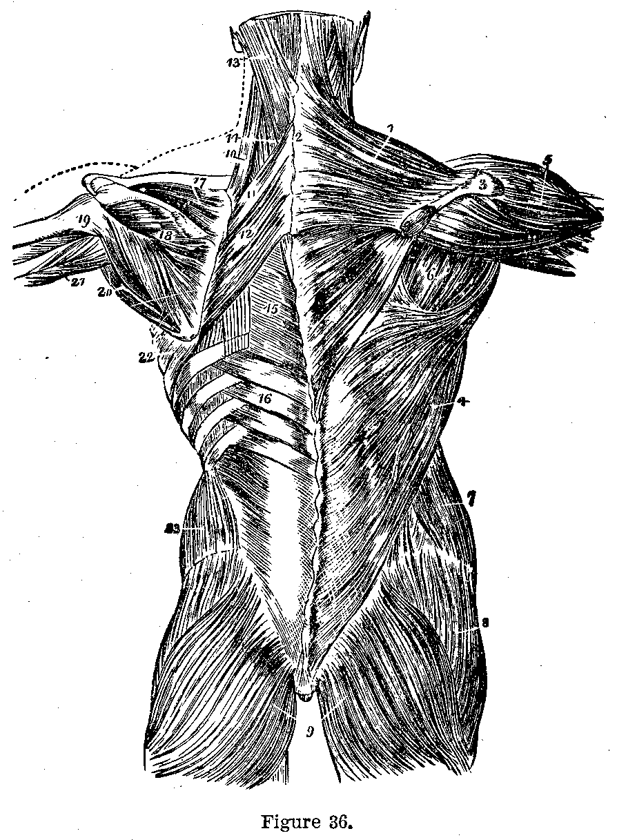
Muscles of the Back.—The most important muscles of the back are the trapezius, which pulls the head back or the shoulder upward or backward and which runs from the occipital bone and the spine as far as the middle of the back, to the shoulder bones; the latissimus dorsi, which draws the arm downward and backward, and which arises from the lower ribs, the lower half of the spine and the haunch bone and is inserted into the arm bone near its head; and the erector spinae which arises from the pelvis and lumbar vertebra and is inserted into all the vertebra above; it maintains the spine erect.
|
|
|
|
|
|
|
Muscles of Thorax.—Concerning the thorax we may mention the intercostal (between the ribs) muscles, external and internal, the external set raising the ribs and the internal set depressing the ribs in respiration.
The Diaphragm.—The diaphragm is a musculo-fibrous partition forming the dome of the abdomen and separating it from the thorax. It is attached to the lower ribs and spinal column and is perforated by the aorta, inferior vena cava and gullet. It is a muscle of respiration and expulsion.
|
|
|
|
|
|
|
The Abdomen.—The abdomen is completed in front and at the sides by a thick wall of muscle which not only aid in protecting the underlying structures but assist in expelling the urine, feces, etc., from, the body. This wall is made of the external oblique muscle which runs from the ribs downward and inward to the pelvic bones and linea alba—the linea alba or white line occupies the midline of the abdomen and is formed by the union of the various muscular structures of the abdominal wall; the internal oblique muscle arises from the ilium and lower fibrous part of the external oblique ( Poupart's ligament), runs upward and inward to be inserted into the linea alba and lower ribs; the transversalis which runs transversely between the brain, spine, ribs and pelvis to the linea alba, and the rectus abdominalis which is situated near the middle line of the body and runs from the ribs to the pelvis.
Breast Muscles.—The pectoralis major and minor muscles form the fleshy masses of the breast. They run from the collarbone, breast-bone and ribs to the caracoid process of the scapula and the humerus, the fibres converging from their origins to their insertions. They draw the shoulder forward and the arm across the chest.
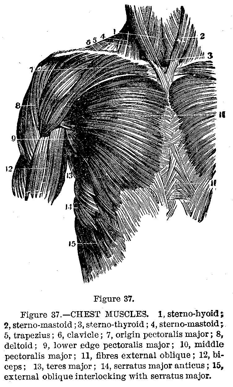
Deltoid Muscle.—The deltoid (Fig. 37) forms the prominence of the shoulder. It arises from the clavicle and scapula, the fibres converging to be inserted into the humerus just above the middle. It raises the arm from the side.
The Biceps.—The biceps forms the prominence on the front of the arm when the forearm is flexed. It arises from the scapula by two heads and is inserted into the upper end of the radius. It flexes the forearm and assists in supinating or turning it over.
|
|
|
|
|
|
|
The Triceps.—The triceps arises from the shoulder blade and the back of the humerus by three heads, and is inserted into the upper end of the ulna. It extends the forearm.
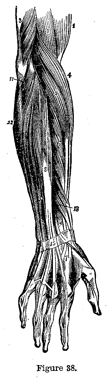
Muscles of Forearm.—The muscles of the forearm are very numerous, and give the forearm and hand a multitude of movements. The muscles which turn the palm downward are called pronators, the most important of these is the pronator radii teres which runs from the inner part of the lower end of the humerus to the radius. The most important supinator or muscle which turns the palm upward (Fig. 38), is the supinator longus which runs from the outer part of the lower end of the humerus to the lower end of the radius.
|
|
|
|
|
|
|
Flexors.—The radial and ulnar flexors (of the wrist) come from the inner part of the arm bone and are inserted into the hand bones. Beneath these muscles lies the flexor sublimus digitorum which divides into four tendons or leaders, one for each finger. These leaders are split so as to give passage to the leaders of the flexor profundus digitorum which are inserted into the ends of the fingers. The thumb is moved by special muscles.
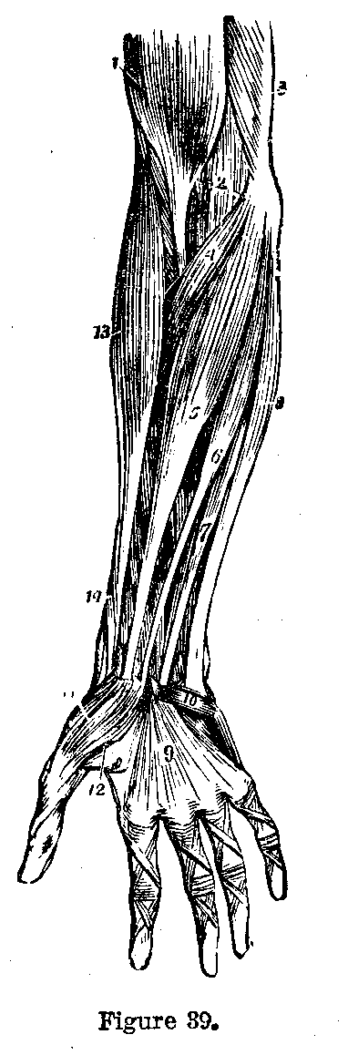
Radial Extensors.—The muscles on the back of the forearm are the longer and shorter radial extensors (of the wrist), which lie behind the long supinator and whose tendons are inserted respectively into the metacarpal bones of the first and second fingers. The ulnar extensor of the wrist lies on the ulnar side of the forearm, and is inserted into the metacarpal bone of the little finger. Between these muscles, in the middle of the forearm (Fig. 39) is the common extensor of the fingers which is inserted by four tendons into the backs of the last two bones of the fingers. The index and little fingers have special extensors. The hand is supplied by a number of short muscles which give it marvelous dexterity.
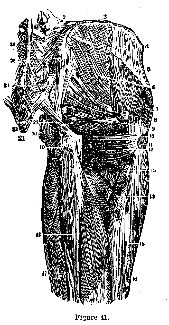
The muscles of the lower extremity consist of those of the hip, thigh, leg and foot. The psoas magnus and the iliacus, the former from the lumbar vertebrae, the latter from the inside of the ilium, are inserted together into the upper part of the femur. They flex the thigh and roll it outward. The buttocks are composed of the three glutei muscles. They arise from the pelvic bones and are inserted into the upper part of the femur. They extend the hips, raise the body from the stooping posture, and hold the trunk on the thigh bones. Partly beneath them lies a group of muscles (Fig. 41), the rotators of the hip; they are the pyriformis, gemelli, the internal and external obturators, and the quadratus femoris.
Thigh Muscles.—The rectus femoris with the vastus externus and internus form the mass of muscle on the front of the thigh. The rectus arises from the ilium, the vastus from the femur; they join to form a common tendon which is attached to the upper end of the tibia. They extend the leg, flex the thigh, and raise the body from the sitting to the standing posture.
The Tailor's Muscle.—The sartorius, the longest muscle in the body, runs from the ilium downward and inward across the thigh to the inner side of the shin bone below the knee. It flexes the thigh and crosses the legs.
|
|
|
|
|
|
|
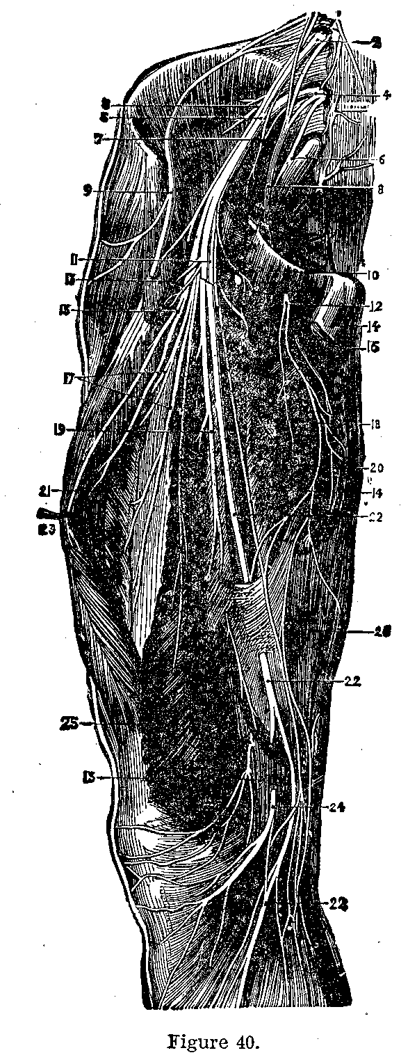
The biceps femoris arises from the ischium and is inserted into the head of the fibula (Fig. 40). The semimembranosus and the semitendinosus take origin from the ischium and are inserted into the inner part of the head of the tibia. They extend the hips, flex the knee, and raise the body from the stooping position.
This page is maintained by
Charles Keith.
Contact:
Send me a message
Last Modified: Monday, 13-May-2013 15:31:46 EDT