|
|
|
|
|
|
|
Bones of the Face.—The bones of the face shown in this plate are the nasal bone, forming the arch of the nose, the malar, which gives prominence to the cheek, the upper jaw, containing the upper teeth, and the lower jaw, containing the under teeth.
The Spinal Column.—That portion of the spinal column noticed in the illustration consists of the cervical vertebrae. Each vertebra is composed of a body, with seven spinous processes projecting from it. The body is perforated by a ring, through which is seen running the spinal cord, giving off nerves between each separate bone. A ring of cartilage is seen inserted between each separate vertebra, the object of which is to prevent any jar reaching the brain when we run, jump, walk or stumble.
Wonderful Structure of the Body.—The human body is the highest form of animal life. It is full of beautiful proportions and divinely symmetrical in shape, form, mould and outline. We look with honest pride and glowing admiration upon the many accomplishments that man has achieved in the world around us. We see his skill displayed in the various arts and sciences, and we look with awe upon the projects of his intellect and reason, the realization of which is but a small question of time! We boast of our ships, our steamboats and our steam cars; we are justly proud of our bridges, our viaducts and the progress of our engineering skill; we grow enthusiastic over our telegraphs, our telephones, our electric lights; we feel a degree of national pride in the achievements and successes of Edison, the wizard of Menlo Park; but where, let us ask, in the whole range of events, the acquirements of arts, the accomplishments of mechanics, the achievements of architecture, the attainments of engineering or the successes and promises of electrical sciences, can we find such another structure as the human body, that curious, yet perfect world of wonders!
Man the Most Complex Body.—It embodies an epitome of the whole universe! Man is more elaborate, more complex, more God-like, than any other living organism; more wonderful, more beautiful, more marvelous, than any work of human ingenuity, conception or construction. Indeed, the mechanism, the skill and the workmanship displayed in the human body is simply perfection itself. In conception, it is divine; in design, perfect; in architecture, grand; in construction, wonderful; in beauty, lovely; in form, symmetrical; in outline, sublime; in strength, great; in arrangements, marvelous; in mobility, transcendent; in adaptability, unexcelled; in fine, when studied in all its parts and their relationship to each other, we are led to exclaim with the Psalmist David, that the human body is "fearfully and wonderfully made."
|
|
|
|
|
|
|
Man the Most Complete Body.—The all-wise Creator, when He first made man, made him perfect. He formed every organ of the body with direct reference to the function to be performed. Every bone, muscle, nerve, organ and tissue formed in the construction of this wondrous organism is made of the right kind of material; is of the proper form and size; placed in the right position to subserve best the purpose for which they were individually and collectively designed, and to perform the peculiar duties assigned to each. We cannot talk with the ears, smell with the eyes, see with the nose, nor walk with the tongue. We cannot think with the lungs, nor breathe with the brain. The stomach was not designed to propel the blood over the system, nor the heart to digest food.
The Complete Organs and Structures.—The muscles which give form and shape to the body would be powerless instruments of movement if devoid of the bones of the skeleton. Thus we see that every organ and structure was formed with direct reference to the accomplishment of a certain definite object. Hence, the bones form a frame work, to protect the delicate organs of mind, respiration, circulation, digestion and excretion, to serve as levers on which the muscles may act to produce motion, and to preserve the form and shape of the body; the muscles, such as we observe in this plate, give form, shape and symmetrical proportions to the body, and produce its varied motions; by means of the brain we think, feel and act; the nerves of the eye take cognizance of external objects, and convey their impressions to the brain; the auditory nerve distinguishes sounds; the olfactory nerve identifies and separates the different odors brought into contact with it, and the sentient nerves of the skin are fully impressed with the touch of external objects, carry the impression of their character and size to the brain, and the motor nerves carry the commands of the will to the muscular system, that the behests of the mind may be obeyed and carried out; the heart receives the impure blood from all parts of the body, and sends it to the lungs to be purified, then receives it back again and forces it with enormous power even unto the most remote and minute part of the system; the arteries and veins are made for the express purpose of conveying the "pabulum of life" from the heart, and to carry the vitiated and poisonous fluid to the heart; the lungs throw off the carbonic acid in the venous blood and replace it by oxygen; the stomach, by and with the aid of the salivary, biliary, pancreatic and intestinal juices, digests the food and transforms it into blood; the kidneys are designed as filters, to aid in the purification of the blood; thus we observe that the various tissues and organs of the body have each their own especial use in the human economy, and their exact and definite function to perform; and as a result of the sum total of the proper required performance of all these different functions, we have not only harmony and health, but happiness of mind, soul and body as well.
|
|
|
|
|
|
|
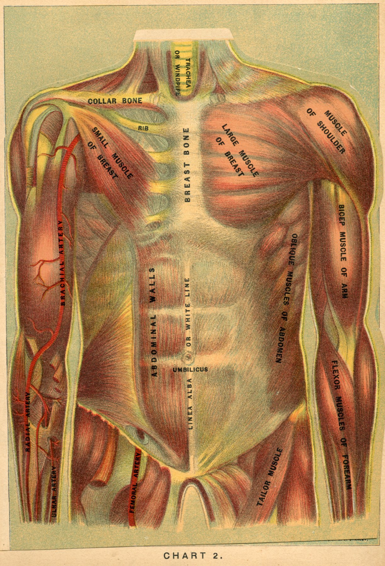
Muscular Arrangement and Blood Supply.—In this exquisite and magnificent colored engraving we have a grand view of the wonderful arrangement of the muscles of the trunk of the human body, together with the muscular arrangement of the arms and likewise their blood supply. The trunk of the body is divided into two compartments— the thorax and abdomen.
The Thorax.—The thorax derives its name from the Greek word thoreo, and signifies "I leap," because the heart leaps in it. It is covered on the front part by large muscles; the pectoralis major, or large muscle of the breast, is observed on the left side of the chest, whilst on the right it is removed and exposes the pectoralis minor, or small muscle of the breast. The dove-tailed muscle observed on each side is the serratus magnus, and is employed in expending and contracting the chest in the act of breathing. The muscles of the chest walls, in a deep inspiration, exert a force equal to lifting a weight of 750 pounds.
Walls of the Abdomen.—The muscular walls of the abdomen are nicely arranged and beautifully adapted to the functions they perform. On the left side we see the large oblique muscle, so named because of the direction its fibres run, and on the right side we observe the rectus muscle, the transverse muscle and internal oblique muscle, all of which are strong, broad muscles, whilst the manner in which they are so scientifically arranged gives additional strength to the abdominal walls, without deteriorating from its great mobility, and at the same time avoiding all pressure of the organs contained within this large cavity. There are ninety-one muscles on each side of the trunk, or one hundred and eighty-two in all, ninety of which are pairs, and two are single.
|
|
|
|
|
|
|
Muscles of the Shoulder.—The large triangular muscle of the shoulder—the deltoid—is one of great strength, as in fact are all the muscles of the arm. If you grasp the arm tightly just above the elbow-joint, and then bend the fore-arm, you will feel the biceps muscle of the arm become firm, hard and prominent; now straighten it again and it becomes relaxed, whilst the muscles on the back of the arm become hard and prominent. The muscles of the fore-arm are the flexors and pronators; that is they flex the arm and turn the palm downward. In each upper extremity or arm there are fifty-three muscles, and we observe here the nicest and most economical method of packing away the muscles that could be improvised, securing strength, giving elegance to its form and shape and facilitating its mobility.
Blood Supply of Arm.—On the right arm we obtain a glimpse of the blood supply of the arm; we see the brachial artery giving off numerous branches, and observe the radial and ulnar arteries doing the same thing; thus securing ample nourishment to preserve the health, strength and beauty of the arm.
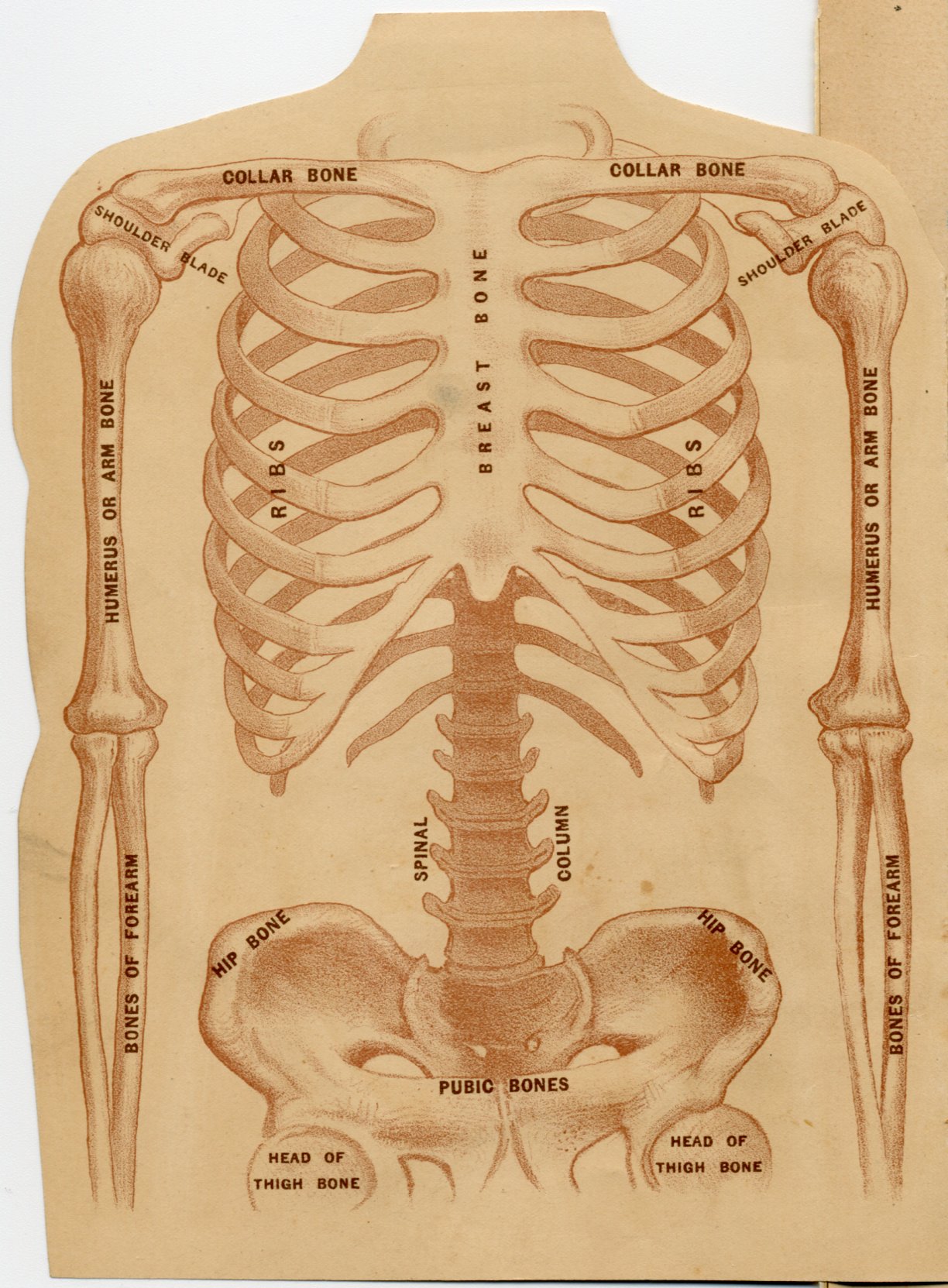
Different Forms of Bones.—On turning over this flap we are brought face to face with a grim looking but useful object—the frame work of the trunk and arms. The skeleton is of a ghastly appearance and emblematic of death; its unsightly look sends a thrill of horror through us and we instinctively recoil from it. Yet it subserves a useful purpose in the human body, and the ugly looking bones, when carefully examined, abound in nice contrivances and ingenious workmanship; whilst each individual bone is designed for the especial duty it has to perform. Hence the bones differ in form; some are long, as in the arms and legs; some are short and thick, giving strength and compactness, as in the lumbar portion of the spine; some are flat, for covering a cavity, as the skull and pelvis, and others used for a special purpose are irregular, as in the hands and feet.
Combined Lightness and Strength.—But notwithstanding this diversity in form, the general plan constantly kept in view by the Divine Architect has been the central idea of combining lightness with the greatest possible degree of strength. The bones of the arms and legs are round, or triangular, and hollow, thus giving with the same weight a greater degree of strength than if solid, besides affording a larger surface for the attachment of muscles.
|
|
|
|
|
|
|
Composition of the Chest.—The chest is composed of bones, cartilages and ligaments. Its natural form is that of a cone diminishing upward; and it affords lodgment for the heart, lungs and large blood-vessels. Its walls are formed posteriorly by the seven dorsal bones of the spinal column, and the ribs as far as the angle, the sides by the body of the ribs, and front by the ribs, the costal cartilages and the breast bone.
The Ribs.—The ribs are twenty-four in number, arranged in pairs, twelve on each side of the chest. At the back they are fastened to the spine, and in front the seven upper pairs are tied by cartilages to the breast bone, three are fastened to each other and the cartilage above, and two, the floating ribs, are loose. The long slender ribs give lightness; their arched form confers strength, and the cartilages impact elasticity; thus the three most essential pre-requisites of the chest for the protection of the delicate organs contained within this cavity are secured, whilst the freest motion in respiration is ensured.
The Pelvis.—The pelvis is an irregular-shaped basin, formed by the hip bones and the pubic bones in front. In the upper and back part is the foot of the spinal column, consisting of a wedge-shaped bone called the sacrum. It is observed firmly planted between the wide spreading hip bones of the pelvis, like the key-stone of an arch, and gives a strong support to the burden above.
The Spinal Column.—The spinal column, the lumbar portion of which is here seen, consists of twenty-four bones, between which are placed pads of cartilage. Such is the elasticity of these cushions of cartilage, that, though they become condensed through the day, making us shorter in the evening than in the morning, they resume their normal thickness while we are lying in bed at night. The perfection in the architecture of the spine surpasses belief; its various uses seem a bundle of contradictions.
Bones of the Spinal Column.—The twenty-four bones of which it consists are so stiffly locked together as to form a chain that will bear and support the heaviest of burdens, yet so flexible that it will bend like India rubber; within this wondrous column hides a delicate nerve that would thrill at the gentlest touch, yet so securely does it rest in its bony couch that it feels not the slightest jar or shock; and resting upon this remarkable pillar of bones is borne the brain without a tremor or a fear of danger; to it are found clinging the vital organs of the chest and abdomen, secure in the protection it affords.
The Shoulder Joint.—The shoulder joint, formed as it is by the shoulder-blade (scapula), collar bone (clavicle), and the arm bone, is most beautifully designed and executed. It comprises a shallow ball and socket-joint, thus affording the freest rotary movements. The shallowness of the socket, however, accounts for the frequent dislocations of this joint; but that is compensated for by the easy, graceful carriage and swing of the arm, which a deeper socket would not permit.
|
|
|
|
|
|
|
The Collar Bone.—The collar bone is fastened at one end to the breast bone and first rib, and at the other end to the shoulder blade. It thus holds the shoulder-joint out from the chest, aids in protecting the important vessels of the axilla, and gives the arm a greater range of freedom, mobility and play.
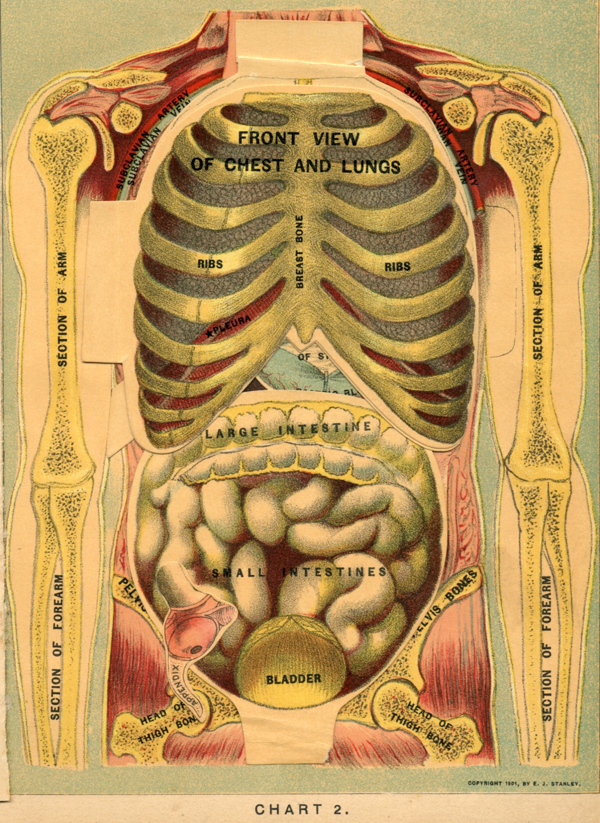
What the Lungs Are.—The lungs! Dense looking objects, and yet how light and buoyant! This beautiful anatomical chart shows us a front view of the chest and lungs, with the lungs enclosed within the bony basket-work of the chest. The lungs are two large, conical bodies, placed one on each side of the chest, and occupy the greater part of its cavity. During life they accurately adapt themselves to the varying dimensions of the chest; for, unhappily, the foibles of fashion very frequently cause restriction of the lungs, by interfering with the resistance and freedom of movement of the ribs, so essential to health, by tight lacing and the barbarous usage of corsets.
Pleura of the Lungs.—In this chart we see also the pleura or the investing membrane of the lungs, and right below it the diaphragm or midriff.
Two Distinct Lungs.—Although the lungs are two in number, as far as their structure is concerned, and are perfectly distinct from each other, having, as we observe in the chart underneath this one, the heart and blood-vessels between them, yet as regards their functions they may be considered the same, since they receive their blood from a single vessel, the pulmonary artery, and the air by one canal, the trachea or wind-pipe, and act in common with each other.
Size and Shape of Lungs.—As will be observed, the lungs are not quite the same size or shape; the right lung, although somewhat shorter and thicker than the left, is the larger and stronger, being divided into three lobes; whilst the left is the smaller and weaker, divided into two lobes only, and hence more frequently subject to disease.
Weight and Shape of Lungs.—The weight of the lungs varies very much; but in general they average about forty-two ounces in the male; thirty-six in the female; the right lung being about two ounces heavier than the left. Each lung is conical in shape, with a broad concave base resting on the convex surface of the midriff, the apex directed upward and extending into the root of the neck about one inch above the level of the first rib.
|
|
|
|
|
|
|
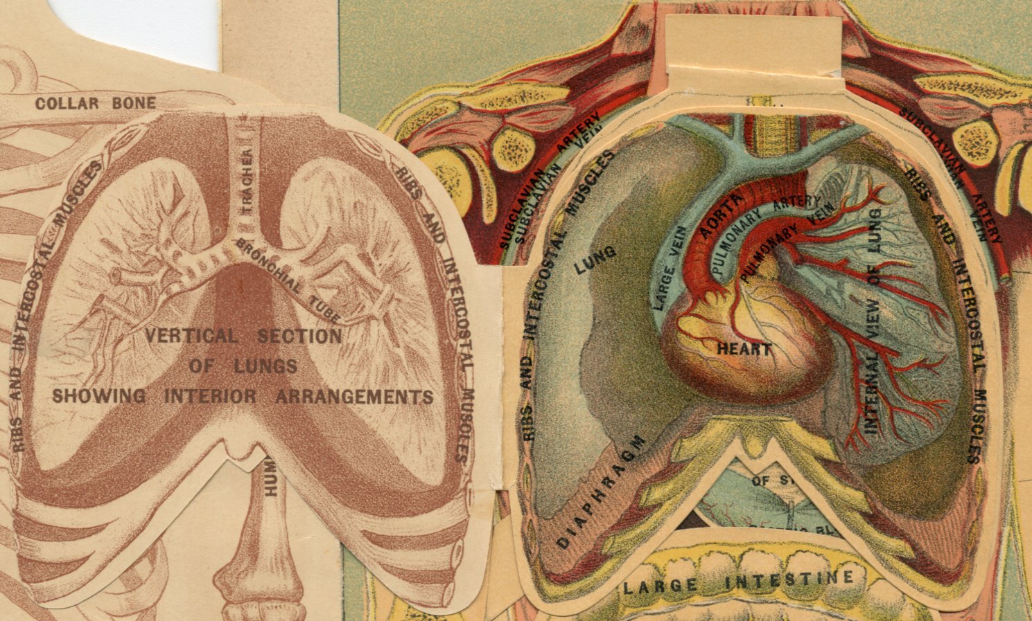
Interior Arrangement of Lungs.—On turning this flap over we find a vertical section of the lungs, showing their interior arrangements. The lower end of the trachea divides, one portion going to each lung. These again subdivide and continue to subdivide in geometrical order, growing smaller and smaller with each division, and extending to every part of the lungs, finally terminating in a cluster of air cells, bound together by cellular tissue and forming a lobule. These lobules vary in size accordingly as they are located on the surface of the lung or deeper in its tissues. Each lobule is separate and distinct from the other, and forms in itself a perfect and independent lung in miniature.
Function of the Lobules.—In this arrangement we see the boundless wisdom of the Creator displayed, for were it not for this wise and perfect provision—one of the very greatest importance in the process of respiration, since it enables each individual lobule to perform its functions independently of the rest—tubercular disease, bronchitis and inflammation of the lungs would not only be incurable, but would prove to be very rapidly fatal.
Lung Air Cells.—Each air cell varies in size from the seventieth to the one two-hundredth part of an inch in diameter. The number of air cells in the two lungs is truly surprising, there being certainly not less than 600,000,000, though according to Dr. Addison's computation there are 1,700,000,000, equivalent to 1,500 square feet of surface on which the process of purifying the blood is constantly and continuously going on in a healthy lung.
Blood-vessels.—On the next flap we have a graphic illustration of the internal arrangements of the blood-vessels of the lungs and bronchial tubes. The pulmonary artery, arising from the right ventricle of the heart, conveys the venous blood to the lungs. It penetrates the lungs and divides and subdivides into branches, which accompany the bronchial tubes and terminate in a dense capillary net-work upon the walls of the air cells, where the blood undergoes that magical change, giving up its poisonous qualities and becoming revivified and healthful.
Pulmonary Veins.—From this net-work of arteries and air cells the radicles of the pulmonary veins arise, and, coalescing into larger and larger branches, at length accompany the arteries and return the blood to the left auricle of the heart in a purified condition. The pulmonary arteries and veins differ from the same vessels in other parts of the body, since the former conveys venous blood, and the latter arterial blood.
|
|
|
|
|
|
|
Breathing.—Respiration, or the act of breathing, consists of the alternate inspiration and expiration of air to and from the lungs; in the process of which the lungs themselves are almost passive instruments, since their contraction and expansion takes place by means of the muscles which surround the chest. The diaphragm or midriff, which, when at rest and the lungs empty, forms a beautiful dome to the abdominal cavity, becomes depressed during the inspiratory process, and presses the walls of the abdomen outward. At the same time the ribs become elevated, thus increasing the size of the chest. Thereupon the elastic lungs expand to occupy the entire space, whilst the current of air, in obedience to a well-known physical law, rushes down the wind-pipe and enters the numerous air-cells, the result of which is inspiration. In expiration the reverse of this takes place. We bend forward, draw the abdominal walls inward, press the diaphragm upward, whilst the ribs are pulled downward. All these acts simultaneously performed decrease the size of the chest, and force or expel the air from the lungs.
Breathing Capacity of Lungs.—The breathing capacity of the lungs bears a close correspondence to the stature of man. For an ordinary-sized man of about five feet eight inches in height, it will be 230 cubic inches, or about one gallon of air, and for each additional inch in stature up to six feet, there will be an increase of eight cubic inches. In a forcible expiration all the air in the lungs is not expelled; there still remains behind 100 cubic inches. Thus with this unexpelled air, the breathing capacity of an ordinary-sized man is about 330 cubic inches, or equivalent to 11 pints of air. Of the 230 cubic inches, 100 can only be forced into the lungs by the exercise of great effort, and is available for emergencies, as striking a heavy blow, or for the purpose of training, as in singing, rowing, running, climbing, etc.; but the extra amount of air always on hand in the lungs is of great value, since it enables the lungs to perforn their functions continuously, even under severe and violent exertions.
Giving Up of Oxygen.—The atmospheric air laden with its life-sustaining properties, oxygen, having passed into the lungs, gives up that vital element and receives in its place the carbonic acid gas, water, and other refuse materials which the blood has picked up in its journey through the body, and which are no longer fitted to circulate in the blood and preserve the vitality of the body. No tonic invigorates so well as a few, deep, full inspirations of pure cold air.
|
|
|
|
|
|
|
Circuit of the Blood.—The blood thus purified passes back to the heart to go on its circuit through the body, every organ of which renews its energy and vigor from the magician's fiery wand, pure, healthy blood; while the air exhaled carries off the impurities.
Change in Color of the Blood.—During this process the blood changes from a dark purple to a bright red. Pure air is the cheapest necessity and the greatest luxury of life. Let it not be the rarest. The relative proportion of the respirations to the pulsations of the heart is about 1 to 4-1/2 or 5; and the quantity of air required to keep the blood pure is very great. Indeed, respiration is the falling weight, the bent spring, which keeps the clock of life in motion; the inspirations and expirations are the strokes of the pendulum which regulates it.
Delicacy of the Organs.—The perfection of the organs which carries on this stupendous office challenges our warmest admiration. So delicately are they arranged that the slightest pressure will cause intense pain, yet tons of air surge to and fro through their intricate passages, and bathe their innumerable cells without our knowledge, so to speak, of its coming and going. We annually perform over 8,400,000 acts of breathing, inhale over 150, 000 feet of air, and purify nearly 4,000 tons of blood! This gigantic and unburdensome process goes on constantly, never wearying or worrying us when in robust health, and we are struck dumbfounded with amazement when the cold calculations of science reveal to us its magnitude and marvelousness.
Second Use of Breathing.—Nor is this stupendousness all. Nature dislikes a waste of energy. In addition to and by a wise adaptation and economy, the process of respiration is made to subserve a second use no less important than that of purifying the blood—the power of speech. The exhaled air, laden though it may be with the human detritus and off-scourings of the body, in passing through the vocal organs can be transformed into prayers of faith, songs of hope and words of good cheer, kindly encouragement and expressions of love!
What the Blood Is.—The blood—the pabulum of life—has not inaptly been termed "Liquid Flesh." But it is more than that, since it contains the materials so essential and so requisite for the building up and repair of every organ and tissue of which the body is composed. The blood is the liquid by means of which the circulation in the body is carried on; it permeates every nook and corner of the system, and is composed of a thin, colorless fluid, the plasma, filled with red disks, so small, flat and thin that it requires 3,500, placed side by side, to measure one inch, and no less than 18,000, placed one upon the other, to make a column one inch in height. These disks are continually forming and as constantly dying.
|
|
|
|
|
|
|
Coagulation of the Blood.—According to Dr. Draper, of New York, 20, 000,000 die at a single breath! Blood when exposed to the air coagulates, and the value of this peculiar yet intrinsic property cannot be overestimated. When an artery is ruptured bleeding takes place, the blood coagulates and forms a plug, thus preventing further hemorrhage. Thus we observe with what Divine foresight and wisdom, not only the wants of the body are provided for, but also the accidents to which it is liable.
Size, Shape and Location of the Heart.—In this beautiful anatomical chart we obtain an accurate idea of the relative size, shape and position of that wonderful engine, the heart, whose tireless efforts to keep the wheels of life in motion are truly surprising, and fill us with amazement at the prodigious work it daily performs. The heart is an irregular, pear-shaped, hollow, muscular organ, placed obliquely in the lower and front part of the chest, between the two lungs and inclining to the left of the centre. The base is directed toward the spine and corresponds with the fourth and fifth dorsal spinal bone, while the apex points between the cartilages of the fifth and sixth ribs on the left side. In this illustration the pericardium, or loose sac in which the heart is enclosed, is removed, and we see the coronary artery with its branches distributed over the outer surface of the complex and restless organ.
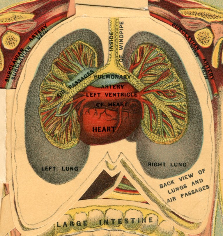
Heart a Double Organ.—On looking at the heart one would think it was a single, solid organ. It is not, however, but a double organ, divided into four compartments; the two upper ones, from their supposed resemblance to a dog's ear, are called auricles, and the lower ones, from resembling a little stomach, are called ventricles. The auricle and ventricle on each side communicate with one another, but the right and left halves of the heart are each separate and distinct organs, and perform different functions—the right side propels the dark, vitiated and impure blood, whilst the left deals with the bright crimson, life-giving and life-sustaining blood.
Use of the Auricles.—The auricles serve as reservoirs to receive the blood—the right, as it comes dark and foul from its tour of the body; the left, as it filters bright and pure from the oxygenated forest of the lungs—and to furnish it to the ventricles as they need it. This is graphically shown on the chart, the large blue vein, formed by the jugular and subclavian veins, is seen descending downward and emptying into the right auricle; the red pulmonary vein, formed by the coalescing of its numerous branches, conveying rich, pure blood from the lungs and depositing it in the left auricle. Corresponding to the lightness of the work they perform, the walls of the auricles are comparatively thin and weak.
|
|
|
|
|
|
|
Ventricles of the Heart.—The walls of the left ventricle, which propels the blood to the remotest corners of the human frame, are correspondingly thicker and stronger than those of the right, which forces the blood to the lungs only. Arising from the right ventricle is seen the blue pulmonary artery, conveying its foul, poisonous, vitiated and venous stream to the lungs, while from the left ventricle is observed the large main artery of the circulatory system—the aorta—from the arch of which arise the right and left carotid arteries.
Changes in the Human Body.—The human body is in a constant state of change. In the midst of life there is death. The blood disks die and new ones are born into life. Every act of life is destructive as well as constructive. Not a thought can be evolved but numerous brain cells die; not a wink of the eye, a smell of a lovely rose, nor a muscular movement, but results in the death of some part of the machinery involved. Every process of life is a process of death. The scales of the epidermis are constantly falling off and being replaced by fresh cells from beneath, and it is on the continuance of this interchange that our life, health and vigor depends. The more rapidly this change goes on, and fresh, vigorous, healthy tissues take the place of the old lifeless ones, the more elasticity, buoyancy and strength we possess—the more healthy and robust we become.
Work of the Heart.—No slave ever performed his work more patiently than the heart. Its quivering task is essential to life and health. It is the fountain from whence the spirit flows, and on the faithful performance of its functions every part of the body depends for the warm stream of life, motion and vigor which it unstintingly furnishes. The ancients believed the heart to be the seat of love. Within its walls were located all that was pure, true, good and noble, as well as the evil passions of the soul. And although modern science has found the seat of mind, reason, consciousness and the mental powers to be located in the brain, and thus robbed the heart of its romance, yet it has revealed wonders connected with this small organ, that certainly eclipse the mysteries associated with it in the past. Pit-a-pat! Pit-a -pat! throbs this marvelous engine, and in response to its constant throbbing the blood bounds along the myriad of tubes, conveying messages of life and health.
|
|
|
|
|
|
|
Constancy of Heart Work.—Our mind cannot stop its beatings; it cannot stop itself; sleep does not interfere with its workings, and our daily labor only strengthens its force and regularity. This wonderful organ throbs on night and day, week in and week out, the year round, with ceaseless, tireless energy. It beats at the rate of 100,000 strokes per day, 40,000,000 per year, and not unfrequently, 2,800,000,000 without a single stoppage. It is the most powerful engine known to science. Its daily work is equal to one-third of that of all the muscles of the body. If it should expend its entire force in lifting its own weight vertically, it would rise 20,000 feet in an hour. The greatest exploit ever accomplished by a locomotive was to lift itself through less than one-eighth of that distance. Vast and constant as is this perpetual throbbing, so perfect is the machinery with which it is carried on, that there are those who do not even know where the heart lies until disease or accident reveal its location.
Vitality of the Heart.—Its vitality is as amazing as its strength. While life exists this tireless organ never stops. In disease, as long as a flutter of this wondrous organ exists, we know the spark of life has not altogether vanished, and new hope is begotten that health may be restored. During such long lives as we sometimes see, the heart has propelled no less than 500,000 tons of blood; and yet, during all this patient, unfaltering and unflinching labor, it has repaired itself as the waste has occurred.
Heart Rhythms.—The rhythm of its beats never fails until death breaks into the casket and seizes the ever throbbing pendulum at the command of the great Master Workman, silencing the quivering muscles of the heart and compelling the wheels of life to stand still.
Value of the Plates.—Seeing is believing; nay, it is more, it is knowing and remembering. The mere reading of a statement on any particular subject does not always advance our knowledge of the matter in question. The observation of a fact, or its proper illustration by appropriate diagrams, such as we observe these anatomical charts to be, not only emphasizes the point considered, but aids us in remembering the principal features connected with the functions performed, thus advancing our knowledge of the subject discussed, and educational progress is made.
Quantity and Variety of Foods.—As we have already seen, the human body consists of numerous mechanics or artisans, who are constantly at work repairing and upbuilding the unceasing destruction that is continually going on. If fresh food be not daily supplied, this work would soon cease, and the lamp of life flicker out. To replace this constant waste we require nearly three pounds of solid food, and fully three pounds of liquid food for our daily allowance. But to convert the pent-up energies of bread, meat and vegetables into the tissues of our own mechanism requires a number of differently constructed organs, and these we now desire to draw your attention to in this beautiful chart. The organs consist of the stomach, liver, pancreas and intestines, which comprise the principal organs concerned in the process of digestion.
|
|
|
|
|
|
|
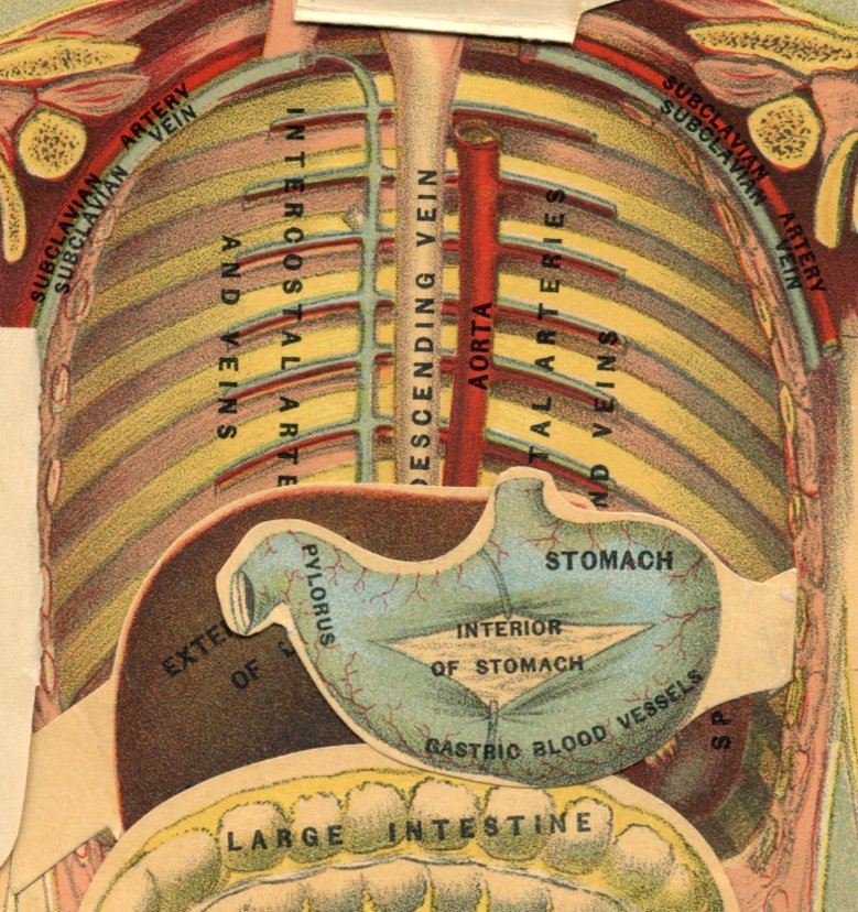
The Stomach.—The stomach is an irregular expansion of the gullet or oesophagus, and is the receptacle which receives the food when swallowed. Its shape has been, not inaptly, likened to the Scotch bagpipe. It will hold about three pints, though it is capable of considerable distension. When moderately filled with food it measures about twelve inches in length by four inches in diameter at its widest end. The walls of the stomach consist of four distinct coats, held together by fine areolar tissue, and are arranged in the following order, from within outward: the mucous, the areolar, the muscular, and the serous. The inner mucous coat is a smooth, soft, rather thick, pulpy membrane, loosely connected with the muscular coat, and secretes the gastric digestive fluid of the stomach.
Fine View of Stomach Coatings.—On turning over the flap we obtain a very fine view of this remarkable membrane. The areolar coat is placed between the muscular and mucous coats, and connects with both. The muscular coat is very thick and stout, and composed of three sets of fibres, the longitudinal, circular and oblique, which form three distinct layers. The outer coat is a thin, smooth, transparent and elastic membrane, derived from the peritoneum, and well lubricated to prevent friction. When the fibres of the muscular wall contract, a peculiar churning movement of the stomach is produced, thus securing the thorough mixing of its contents, that every particle may come into contact with the solvent properties of the gastric juice.
The Pyloric Gate.—At the smaller end the muscular fibres contract and form a gateway—the pylorus, as it is called—which carefully guards the exit from the stomach, and allows no food to escape until properly prepared. The gastric blood-vessels are seen distributing themselves over the outer surface of the organ, thus ensuring its nutrition and repair.
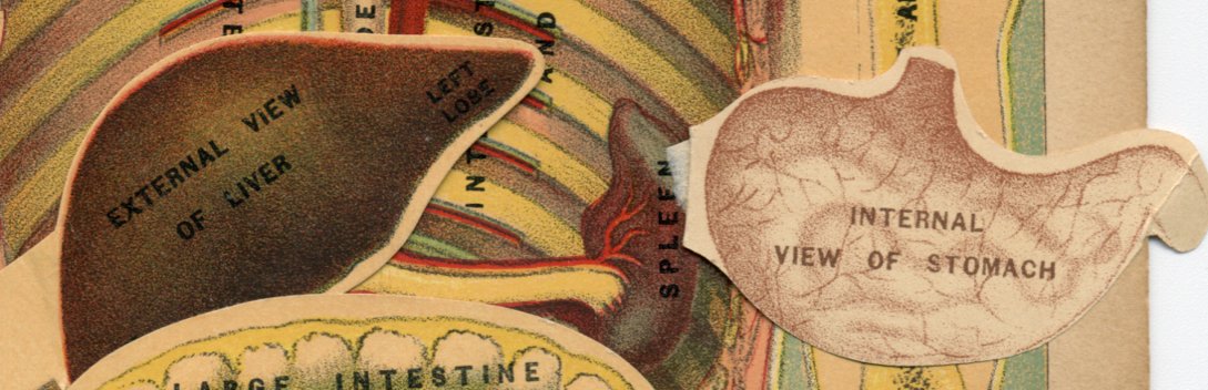
The Liver.—The liver is the secreting organ by which the bile is formed. It is situated on the right side below the diaphragm, and is of a reddish-brown color. It is irregular in form, being convex on the upper surface, irregularly concave behind, very thin in front, and weighs about four pounds. It is, therefore, seen to be the largest organ or gland in the body. It is divided into two lobes, the right and the left, the former being by far the larger. On turning the flap over, we see how intricately it is arranged internally.
|
|
|
|
|
|
|
Blood-Vessels of the Liver.—The blood-vessels of the liver are the hepatic artery and veins, and the portal vein; the lymphatic vessels are numerous, and the nerves are supplied from the pneumogastric, the phrenic and the hepatic plexus. The liver, therefore, receives two kinds of blood: the arterial, by means of the hepatic artery, and the venous, from the portal vein, from which the bile is principally formed. The bile is a dark, golden fluid, of extremely bitter taste, of which three pounds is secreted daily. When not used in digestion it is stored away in the gall-bladder, a fine view of the location of which we have in this chart. The action of the bile on food, though not fully understood, is necessary for perfect digestion.
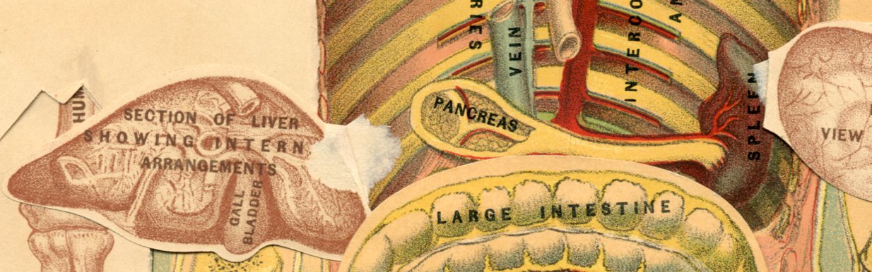
The Pancreas, or "Sweetbread."—The pancreas, or "sweetbread," is a single glandular organ, situated transversely across the upper and back part of the abdomen, on a level with the last dorsal spinal bone. It is of an irregular, elongated form, from six to eight inches in length, an inch and a half in breadth, and from a half to one inch in thickness. It secretes about seven ounces daily of a slightly alkaline fluid containing an organic principle—pancreatin, which has the property of changing the starchy food into sugar. Whilst it has this power, yet its chief work in the digestive process seems to be the breaking up of the fat globules into myriads of minute particles which mix freely with water, and thereby promote their absorption by the lacteals.
The Intestines.—The next chart shows us the manner in which the intestines are arranged in the abdominal cavity. The entire intestinal canal is about thirty feet in length, and is divided into two portions—the small intestines, and the large intestines; these again are each subdivided into three different portions. Of the large intestines, the transverse portion is laid open, showing the internal arrangements. A section of the bladder is seen on this chart.
Machinery of Digestion.—From the number and differently formed structures which constitute the digestive organs, it will be observed that that function is a very highly complex process. If the food were thrown directly into the circulating fluid, it could not be used for the purpose of nutrition. It requires for its transformation into blood, bone and muscle, a series of complex machinery, each part of which is specially designed for the particular part it plays in this wonderful and complicated process.
|
|
|
|
|
|
|
Use of Mouth and Teeth.—The mechanical part, which, although not shown in this chart, may be carefully studied in the chart giving the different views of the head, is performed by the mouth and teeth, and the pulverized food is subjected to the action of the saliva. The lubricated morsel of food is now gathered into a ball and conveyed to the back of the mouth by the muscles of the cheek and tongue. On its arrival here, the soft palate lifts upward and. closes the posterior nasal openings; the epiglottis shuts down over the trachea or wind-pipe, forming a bridge over which the food passes, thus preventing it from falling into the respiratory track.
Duty of the Throat.—The muscular bands of the throat now grasp it and pass it down the gullet into the stomach, beyond our control. Here it comes into contact with the gastric juice, undergoes the churning motion of the stomach, is guarded over by the pylorus, thoroughly saturated and mixed before entering into the intestinal track, where it is subjected to the action of the bile, the pancreatic juice and the intestinal fluid, each with its special duty to perform.
Nature's Treasures Open to Man.—All this is a very complicated and diversified process, the necessity for which can only be explained upon the hypothesis that Nature, in her exhaustless munificence, has opened her proud domains, and poured forth to man the treasures of every land, and every sea for food; the cornfields wave their golden grain for him; the wheat, rye, oats, corn, maize, rice, each different, yet highly nutritious and sufficing; the palm, the date, the banana, the fig, the pineapple, spread out a delicious harvest on the air; the luscious apple, pear, peach, plum, cherry, tempt his ready hand; the potato, the beet, the turnip, the tomato, the cabbage, the pea, the cauliflower, and a thousand other good things, incite his appetite, whilst to this feast is added the flesh of birds, of oxen, of sheep, of swine and of fish; that before the waving wheat and corn, the flesh of other animals, the fruits and farinaceous foods, the running water, the luscious oyster and fish, etc., can be transformed into the refined and spiritual organization of man, it must be thoroughly prepared by the several steps in the digestive process—then, and only then, is it permitted to enter into and commingle with the highly complex, nutritious and life-sustaining fluid, the blood.
Great Value and Beauty of the Plate.—We can understand much of this wonderful process. We have looked into the stomach, watched its peculiar actions and traced its various steps, from which the scientist is capable, in his laboratory of knives, mortars, baths, chemicals and filters, of imitating many of the operations of digestion; but just at the moment he thinks himself most successful, he is compelled to pause. At the threshold of that "one step more," which Fontenelle required, "and he would surprise nature herself," he stops, and very wisely, without concealment of his designs, admires, then wonders, and finally worships with all the reverence of his soul.
|
|
|
|
|
|
|
Transformation of Food Into Flesh.—How strange this is—the transformation of food into human flesh, into human thoughts! We eat a meal; it is composed of meat, bread, vegetables and liquids. The more solid part is ground by the teeth, mixed with the different juices, dissolved, changed, organized and is swept through the body in the circulation of the blood. Each organ seizes its own particular food as it passes. Within the cells of the various tissues it is transformed into the soft, sensitive brain, or the hard callous bone; here into the nerve of sight, there into gristle or tendon; here briny tears are formed, there the bland saliva in the stomach, acid juice; in the skin, acrid perspiration; bile for digestion, oil for the hair, nails for the fingers, muscle for the strong arm of toil, and flesh and fat to give shape, form and beauty to the face.
Wonderful it all is.—Wonderful! Within us is an Almighty Architect, who superintends a thousand skilled laborers, that make a way which puzzles human comprehension; here a fibre of muscle; there a filament of nerve; here constructing a bone; there uniting a tendon—fashioning each with the most scrupulous care and unerring nicety. Without the buzz of a saw, or the sound of a hammer; without the slightest compression, or the least particle of noise; with a regularity, certainty and exactness, the glorious temple of man, in the image of his Creator, goes up and up, day by day, skilfully put together by these noiseless, tireless and expert workmen.
The chart brings into view the location and form of that wonderful little organ known as the Vermiform (wormlike) Appendix (appendage). It is an appendage of the Caecum, or lower bowel. Its uses as a bowel appendage have never been established. It occurs in other animals besides man. Notwithstanding its diminutive size and uselessness as an organ it is the seat of that most painful and dangerous disease called Appendicitis, which was formerly attributed to the presence of some foreign body, as a grape-seed, lodged within it. But it has been found inflammation of the Appendage may arise from numerous other causes. Many doctors insist that a surgical operation, that is the entire removal of it with knife, is the proper curative agent, especially in acute cases. But others insist that the knife is too frequently used, and that the disease, if taken in time, is quite surely curable by absorption.
|
|
|
|
|
|
|
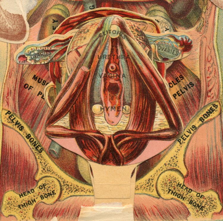
These female organs show further the perfection which anatomical plate printing has attained.
Clitoris.—This small organ, it will be seen, is situated at the upper part of the vulva, or outside parts of the female generative system, and is usually concealed by the lips of the pudenda. It performs a function during sexual intercourse similar to that performed by the penis of the male.
Urethra.—This highly useful organ, common to both sexes, is, as will be seen by the plate, the canal, or medium, by means of which the urine is carried from the bladder to be voided. It is a delicately lined organ, furnished with retentive valves, and therefore susceptible to a variety of diseases.
Vagina.—This word implies a sheath, and is applied to the canal which leads from the uterus (womb) to the external organs of generation in the female sex. Commonly, it implies such external organs, or organ, as depicted in the plate.
Hymen.—The mucous membrane, or virginal membrane, at the entrance of the female sexual organ, or vagina.
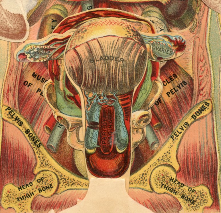
The function and form of the bladder are familiarly known. It is the recipient of the kidney secretions, and contains them till voided through the urinary canal. It is of tough, elastic structure, guarded at the exit by a contractile valve, by means of which the urine can be retained until the quantity becomes excessive. The plate brings out the entire urinal tract, from the bladder to the vagina, and presents a fine and useful anatomical and physiological study.
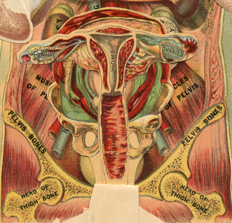
The Womb.—The plate beautifully and effectively illustrates the location and formation of the womb, that wonderful organ which performs the function of parturition, and which is so constructed as to assist in all the necessary efforts of birth. Its structure is elastic and strong, and it expands readily to accommodate the growth of the child (foetus). While this is true exteriorly, its inner parts are rather delicately lined, and subject to a variety of painful diseases, generally designated as "Diseases of the Womb."
|
|
|
|
|
|
|
The Ovaries.—The organs are situated contiguously to the womb. They signify eggs from their shape, and they are the parts which the male semen acts upon to produce the phenomenon of pregnancy. Their enlargement by inflammation and their passage down the fallopian tubes, once a month during the middle period of female life, produces the condition familiarly known as menstruation. The plate also affords another view of the vagina.
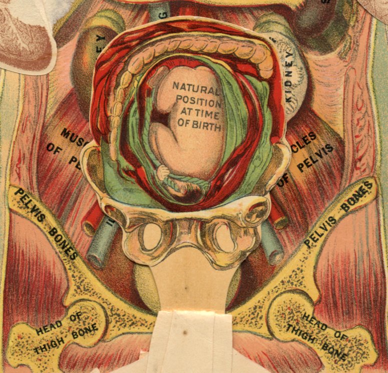
This beautiful and effective plate shows the natural position of the child at the time of birth. It is technically called the presentment of the foetus for birth. Of presentments there are many varieties, whose study is most interesting to the obstetrician. Some of them give rise to very difficult and dangerous delivery. When the presentment is natural, as in the figure, the comfort of the mother is increased and the doctor's anxiety is much allayed.
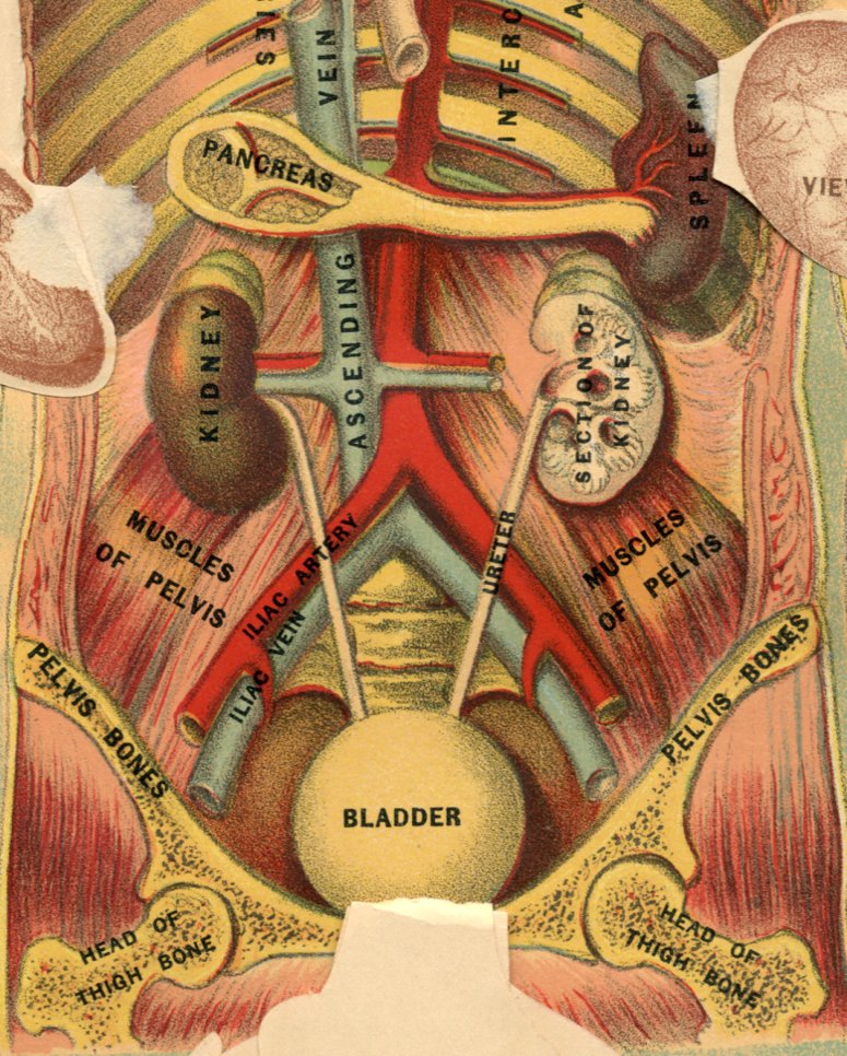
Blood-Vessels of the Body.—The blood-vessels of the human body consist of the heart, arteries, veins and capillaries. The heart and its wonders we have already referred to. In this magnificent chart we are enabled to form some idea of the larger blood-vessels. We see the main arterial tube of the body—the aorta—from a point where it unites with the arch of the aorta; and in its descent downward along the spinal column it gives off numerous branches.
The Arteries.—Opposite the fourth lumbar vertebra it is seen to split in two, and these divisions are called, from their position, the right and left iliac arteries. These are seen to divide again into the internal and external iliac arteries, the former of which is distributed to the walls and viscera contained in the pelvis, then proceeding to the lower limbs after sending two important branches to the abdominal walls. The arch of the aorta gives off the innominate artery, which divides into the right carotid and right subclavian arteries; the left carotid and left subclavian spring direct from the arch of the aorta. Each carotid artery divides into the external and internal carotid arteries, the former being distributed to the external parts of the face and head; the latter supplies the brain and internal parts of the cranium. The subclavian arteries supply the upper extremities with blood.
|
|
|
|
|
|
|
Intercostal Arteries.—The intercostal arteries and veins are beautifully illustrated in the chart. The veins return the blood to the heart, The large ascending and descending venae cavae are seen in this illustration.
Meaning of Artery.—From the fact that at death the arteries are empty, the ancients believed them to contain air, whence their name, derived from aer, air, and tereo, I keep, which literally means, air ducts.
The Spleen.—The spleen is a spongy organ, of a livid color, oval in figure and situated in the left upper part of the abdomen and immediately behind the stomach. Its weight varies from four to ten ounces. It is largely composed of cells, but its function is little understood, though from its position it is believed to be in some way useful to the stomach during the process of digestion.
The Kidneys.—The kidneys are two glandular bodies, having for their functions the secretion of urine. The form of the kidney resembles a French bean; its average length being from four to four and a half inches, two inches in breadth and one in thickness. The two kidneys are situated one on each side of the spine in the lumbar region, opposite the last two dorsal and two first lumbar vertebrae; they are of a brownish-red color, flattened from before backward, and grooved on the interior border for the reception of the great vessels.
The Veins.—The venal arteries are derived direct from the aorta; and the large veins terminate in the ascending large vein. On the right kidney is seen the super-renal capsule; whilst the left is cut vertically into showing the uriniferous tubes, much convoluted and inosculating with each other. The ureter is seen arising from the pelvis of the kidney, descending in an oblique manner to the bladder. These wonderful little organs appear to act as filters, and thus assist to keep the vital stream of life in as pure and as healthy a condition as possible.
The Bladder.—The bladder is a thin membranous bag, which serves as a receiver of the urine secreted by the kidneys, and which remains there until voided by urination through the urethra.
Bone Sections.—The sections of the bones show their cancellated appearance, which combines lightness with strength.
This page is maintained by
Charles Keith.
Contact:
Send me a message
Last Modified: Monday, 13-May-2013 15:31:46 EDT